When bioarchaeologists excavate human remains, they are sometimes faced with burials that can be somewhat complicated. A ‘perfect’ burial for bioarchaeological analyses would contain a complete set of bones that are well preserved. Some archaeological sites, however, are of such great significance that we even work with poorly preserved and incomplete burials, using unconventional methods that allow us to overcome these preservation limitations. This was proven by our research team whilst studying the Bronze Age cemetery of Deh Dumen, located in the Zagros Mountains in Iran.
A significant graveyard in Iran
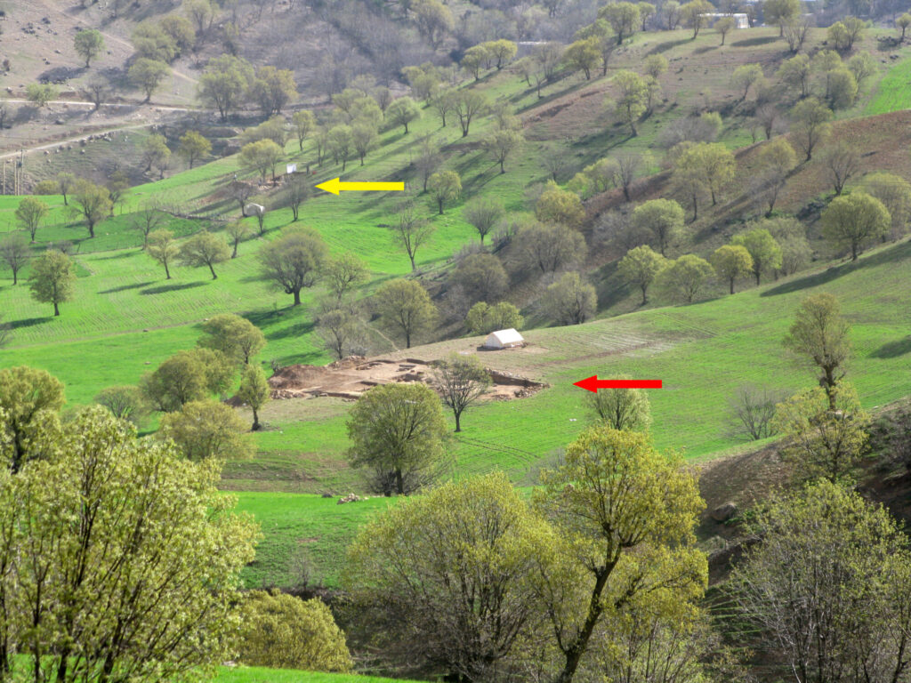
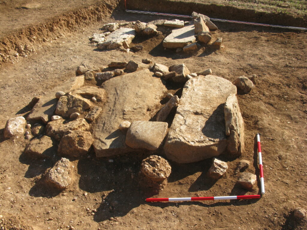
The Bronze Age cemetery of Deh Dumen is an archaeologically significant graveyard for the history of Iran, and the Near East more broadly. The site has yielded important grave goods, such as pottery and metal items, that suggest Deh Dumen played a role in connecting ancient people from the east and west, who settled there and used the graveyard to bury their dead ca. 3000 − 1500 BC. Reza Naseri, an archaeologist from the University of Zabol, has been the director of excavations at Deh Dumen over several seasons. He unearthed graves containing several assemblages of adult human skeletal remains. These multiple burials are fascinating as they likely indicate some form of cultural meaning behind placing several individuals in one grave. What they also result in centuries later is the mixing up (commingling) of bones; throw in inconsistent preservation, including damaged ends of long bones, missing bones and fragmentation — all of which can be found at Deh Dumen — and you have a site which is extremely challenging for bioarchaeologists. Can we learn anything about the lifestyles of these people from such limited evidence?
©R. Naseri, under CC BY-NC-SA 4.0
Unconventional methods for bioarchaeological research in action
Back in 2016, I connected with Arkadiusz Sołtysiak, a bioarchaeologist from the University of Warsaw, to study human remains from the site. We started a long-term project using bone histology to reconstruct the behaviour of ancient Iranian societies. The Deh Dumen cemetery is one of the first sites we have successfully analysed — the results were recently published in the journal Archaeometry.
Deep into the structures
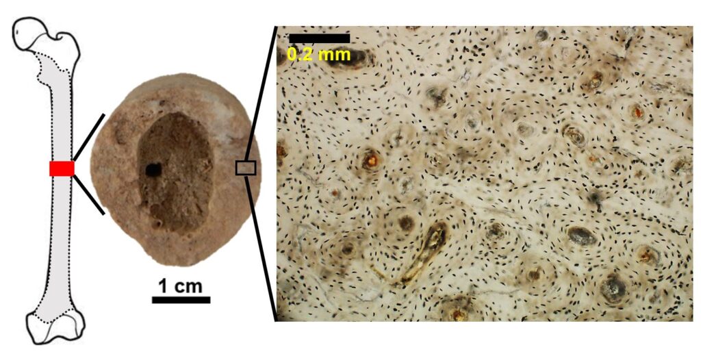
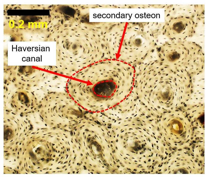
Histology is the study of microscopic structures that make up biological tissues. It is not a novel technique as it has been around since at least the 18th century, when Marie François Xavier Bichat first recognised different types of tissues that make up human organs. Classic thin section histology involves extracting a small piece of tissue and processing it down to a very thin slice (less than 0.1 mm), so that light can be shone through it under a microscope, to identify cells and the organisation of tissue. Bioarchaeologists can apply histology to archaeological remains, but it is not a routinely conducted procedure due to its inherent invasiveness.
Working with irreplaceable archaeological human specimens calls for non-destructive approaches, ideally a visual assessment of gross anatomy and/or non-invasive X-ray microtomography scanning. In cases where thin sectioning is approved on archaeological specimens, 2D images of cortical bone can be examined for geometric properties of bone structures that are a result of remodelling — a process which adapts our bones to various factors including sex, age, disease, and biomechanical loading. Bone histology combined with adult bone shape and form tells us about the loading history of the bone itself, and therefore that of its owner when alive.
Incomplete bones of Deh Dumen
The poor condition of the Deh Dumen specimens meant bone histology was an ideal tool to tackle behaviour questions. For this purpose, we used femur bones with well-preserved shaft segments. We were able to collect some data about the size and shape of the femur midshaft (cross-sectional geometry) and the underlying histology parameters. Our team had estimated that a minimum of 23 individuals would have been represented by the Deh Dumen bones. This gave us a good enough sample size to allow for statistical analyses. Arkadiusz had also previously estimated sex from the femora, finding 13 males and seven females, and three skeletons of indeterminate sex.
Because bone structure varies with age and sex, in addition to lifestyle, we had to somehow account for age despite not having enough skeletal material for its reliable estimation (bioarchaeologists would typically need the teeth, skull and other matched postcranial bones to do this). Histology came in handy yet again, because bone microstructure changes as we age. Bone starts off with primary layers which are replaced with secondary bone that is then repeatedly remodelled. Thus, we were able to categorise the Deh Dumen samples into groups of young adults and old adults on this basis.
©R. Naseri, under CC BY-NC-SA 4.0
Investigating the division of labour by sex
Ethnographic accounts for modern rural Iranian communities often discuss transhumant pastoralism, the mode of subsistence where livestock is seasonally moved up and down mountains. A sex-specific division of labour is very common in these types of communities, where males engage in livestock activities, whereas females are more involved in food production tasks that are more household-bound and sedentary. Because the Deh Dumen site is located within the Zagros Mountains, and other archaeological evidence suggested pastoralism would have been practised there for thousands of years, we hypothesised that the bones of Deh Dumen males should be more robust and experience more remodelling compared to females.
The data were somewhat surprising! When considering the cross-sectional geometry measurements, we found that indeed the male femoral bones were wider and shaped more irregularly compared to females. This supported the sex-specific labour idea, as it indicated males might have received more bending and torsion stimulation from activities involving regular walking up and down the mountains. However, bone remodelling markers (specific indicators used by researchers to understand the process) did not seem to differ much between the sexes. This suggested that behavioural factors shaping the leg bones of Deh Dumen individuals might have differed throughout their life-course. Our bones are predominantly modelled (shape and size of bones) in the first two decades of life, when we are still actively growing to attain an adult skeleton. The long bone cross-sections change quite dramatically during this period, and having biomechanical input is vital. For the reminder of our adulthood, once modelling mostly ceases, we experience remodelling that adapts bone to function in a targeted way, i.e. where it is needed. Thus, it is possible that the young Deh Dumen males and females participated in sex-specific pastoralist tasks, but with age, the line between typically male and female tasks might have become blurred.
© J. Miszkiewicz, under CC BY-NC-SA 4.0 license
Dominant left or right leg?
In the small sample presented here, we also had one individual for whom both the left and right femur were available. This was a great opportunity to test for possible unilateral differences in bone remodelling, assuming walking up and down mountains involves a dominant leg (it might seem counter-intuitive to think that for our lower limbs, but experimental sports science research has shown that most humans do indeed have a dominant leg). We were astonished to find statistically significant differences between the left and right femur in this particular case, as the right leg showed evidence for quicker bone remodelling events. This confirmed our idea that some of these individuals might have practised a transhumant type of pastoralism moving over raised wadis.
We wish we had access to more and better-preserved samples to let us apply more controls to our study, as it is possible our results are being confounded by other aspects including diet. However, the nature of archaeological research is that we have to work with what we have. There are two exciting findings that our study has identified. One is that even commingled and poorly preserved bones can still be meaningfully analysed using unconventional methods, such as histology. We tied histology to behavioural markers, and also age estimation. The second finding is that by combining histology with traditional methods of behavioural analyses (cross-sectional geometry), we can get an insight into different phases (modelling and remodelling) of bone adaptation to behaviour, allowing us to refine the reconstruction of the lifestyles of past people. Our collaboration is ongoing, and soon we will hopefully have more data for other ancient sites in the Near East.
Author:
Justyna Miszkiewicz – skeletal biologist and histologist, currently employed in the University of Queensland. She applies histology to modern and archaeological samples to understand the intricacies of bone remodelling, with implications for reconstructing past human lifeways, as well as understanding bone fragility in humans today. Editor of the Proceedings of the Royal Society of Queensland, and Editorial Board Member of Anthropological Review, and Scientific Reports.
Editing: A.C.
Proofreading: S.A.